CELLS, TISSUES, ORGANS AND ORGAN SYSTEMS
Day 1
The students were asked to look through the unit in the textbook (pages 2-99) and to find topics that they anticipate will be discussed in this unit. They are to use these ideas to create a theme page that includes the following criteria:
| title_page_criterion_1.docx |
Day 2 - The Early Microscope
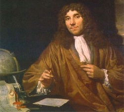
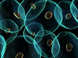
"The father of microscopy, Anton Van Leeuwenhoek of Holland (1632-1723), started as an apprentice in a dry goods store where magnifying glasses were used to count the threads in cloth. Anton van Leeuwenhoek was inspired by the glasses used by drapers to inspect the quality of cloth. He taught himself new methods for grinding and polishing tiny lenses of great curvature which gave magnifications up to 270x diameters, the finest known at that time.
These lenses led to the building of Anton Van Leeuwenhoek's microscopes (for more on the early microscope check out this weblink:http://bit.ly/9RbQtD)considered the first practical microscopes, and the biological discoveries for which he is famous. Anton Van Leeuwenhoek was the first to see and describe bacteria (1674), yeast plants, the teeming life in a drop of water, and the circulation of blood corpuscles in capillaries. During a long life he used his lenses to make pioneer studies on an extraordinary variety of things, both living and non-living, and reported his findings in over a hundred letters to the Royal Society of England and the French Academy." http://inventors.about.com/library/inventors/blleeuwenhoek.htm
The following clips provide a summary of early microscopy and those who were influential in advancing it. The first focusses on Von Leeuwenhoek and the second on Robert Hooke:
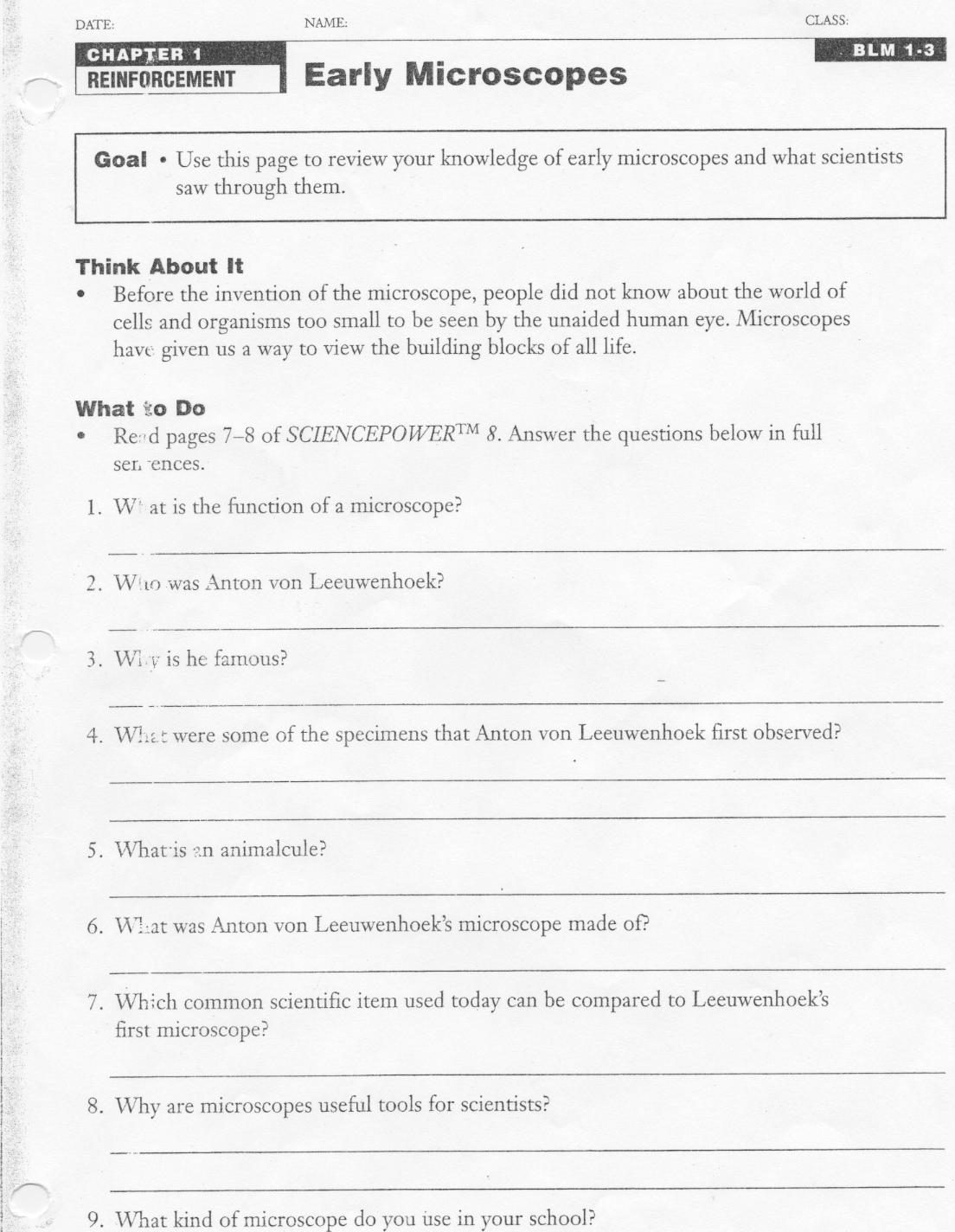
Students were asked to complete the "Early Microscopes" activity sheet.
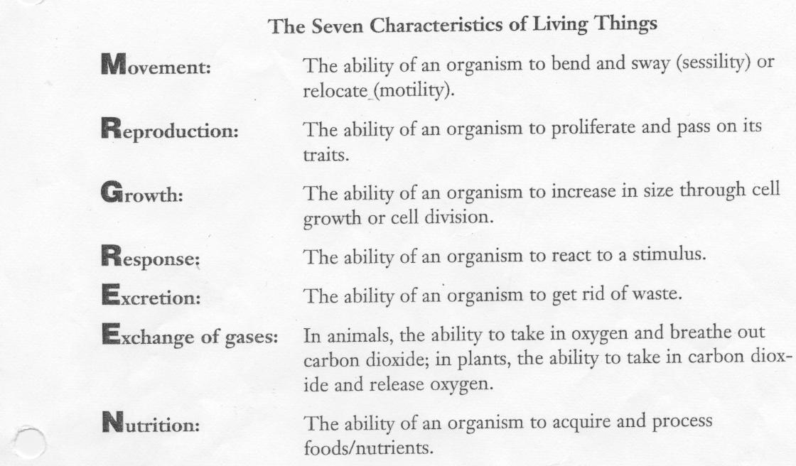
| 7_characteristics_of_living_things.jpg |
| early_microscopes.jpg |
Day 3 - Microscopes Today
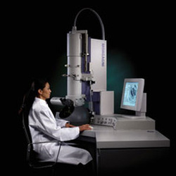
Microscopes have come a long way from being instruments just used to see small objects in the 18th century. The invention of the microscope proved to be a major breakthrough in scientific and medical history. It was possible to look into human blood and see what was going on inside. Doctors and researchers could identify different bacteria and start creating medicines to counter them. It opened up avenues never explored earlier. In short it proved to be a huge boon to us human beings.
Microscopes Today
Microscopes are available in many different types and models. You can get economy ones as well as expensive ones. The best microscopes are made of metal alloys and are long lasting. Microscopes can be used in almost every sphere of life. They are used in schools, hospitals and clinical laboratories. They can be used for metallurgical and industrial work as well.
Types Of Microscopes
Different types of microscopes are used for different purposes. There are four types of biological microscopes that are used to examine tiny matter like blood cells and bacteria. These are the compound, dissection, transmission electron and scanning electron microscopes. Each one can be used for different functions. The compound microscopeallows you to view specimens at a high magnification power. It uses a fluorescent light to illuminate the specimens. The dissection microscope is like a regular compound microscope. It also uses light to illuminate the specimen. It has a lower magnification power and can be used to examine larger organisms by dissecting them. Electron microscopes use electrons instead of light to illuminate the specimens. These have very high magnifying power and resolution. They are used to examine blood and bacteria. They are especially useful in medical research. The digital microscopeis one of the latest innovations. It uses a USB to transfer images to a computer screen. This makes it easier to see the image and manipulate it on a larger screen. It also makes it easier to transmit such images to other scientists in other locations.
Microscopes are used in many locations now. The introduction to microscopes begins in school where they are used for educational purposes. Students conduct experiments by using dissection microscopes to examine small organisms. Microscopes are used in clinical and medical laboratories to examine blood samples to determine the kind of bacteria in them. This helps doctors diagnose their patients correctly and treat them accordingly. Microscopes are used extensively in research laboratories. Scientists are constantly examining different matter and specimens to learn more about them. Thanks to microscopes we discovered different diseases and the cures to many of them. Scientists are still conducting research by using microscopes to find cures and vaccines for many more life threatening diseases. There are microscopes used to examine non-biological specimens as well. They are used to examine and understand the composition of metals, rocks and even fossils. As you can see, microscopes are used in most areas of our lives.
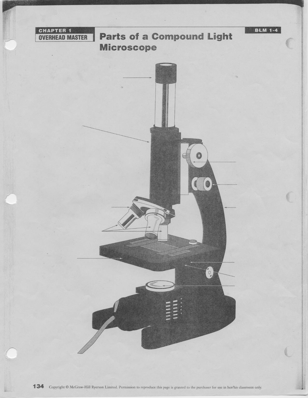
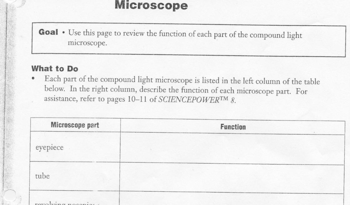
Today the students were asked to identify the location and function of each part of the compound light microscope. They were asked to complete the following activity pages.
The following weblink provides an (easy to read and) indepth explanation of the compound light microscope, how to use it properly and some prepared slides for examination. Enjoy!
http://bit.ly/rql3hZ
| the_parts_of_a_compound_light_microscope.jpg |
| the_compound_light_microscope.jpg |
The following clip outlines the locations of the parts of the compound light microscope and also how to use it safely.
